Dr. Rice has joined Beacon Orthopedics and Sports Medicine
Congratulations Dr. Rice: 2025 Cincinnati Magazine Top Doctor
ACL Reconstruction
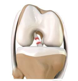
Anterior cruciate ligament (ACL) reconstruction is surgery to reconstruct the torn ligament of your knee with a tissue graft. Anterior cruciate ligament is one of the four major ligaments of the knee that connects the femur (thigh bone) to the tibia (shin bone) and helps stabilize the knee joint. Ligaments are tough, non-stretchable fibers that hold your bones together. Anterior cruciate ligament prevents excessive forward movement of the lower leg bone (tibia) in relation to the thigh bone (femur) as well as limits rotational movements of the knee.
Anterior cruciate ligament injury is a common knee ligament injury. If you have injured your anterior cruciate ligament, surgery may be needed to regain full function of your knee.
Causes of ACL injury
An ACL injury most commonly occurs during sports that involve twisting or overextending your knee. The ACL can be injured in several ways:
- Sudden directional change in motion
- Slowing down while running
- Improper landing from a jump
- Direct blow to the side of your knee, such as during a football tackle
Symptoms
When you injure your ACL, you might hear a loud "pop" sound and experience a buckling of the knee. Within a few hours of an ACL injury, your knee may swell due to the bleeding from blood vessels within the torn ligament. You may also notice instability of the knee, especially when trying to change direction of the knee, during movement.
Diagnosis
An ACL injury can be diagnosed with a thorough physical examination of the knee and diagnostic tests such as X-rays, MRI scans and arthroscopy. X-rays may be needed to rule out any fractures. In addition, your doctor will often perform the Lachman’s test to see if the ACL is intact. During a Lachman’s test, knees with a torn ACL may show increased forward movement of the tibia compared to a healthy knee. Pivot shift test is another test to assess ACL tear. During the pivot shift test, if the ACL is torn the tibia will move forward on complete straightening of the knee and as the knee bends past 30° the tibia shifts back into correct place in relation to the femur.
Treatment
Treatment options include both non-surgical and surgical methods. If the overall stability of the knee is intact, non-surgical treatment is recommended. Non-surgical treatment consists of rest, ice, compression, and elevation (RICE protocol); all assist in controlling pain and swelling. Physical therapy aims to improve knee motion and strength, and resestablish proprioception, or the position sense of the joint in space.A knee brace may be helpful to stablizethe knee with side-to-side or rotational movement. If left untreated or undertreated, an improperly rehabbed ACL injury can result in weakness, persistent gross instability or microinstability, and subsequently can lead to early arthritis of the knee.
An ACL reconstruction surgery is considered for active adult patients, who participate in pivoting sports and require knee stability for work.The goal of ACL reconstruction is to tighten the knee joint and restore its stability. It also helps you avoid further injury and allows you to return to your sport.
ACL Reconstruction Surgery
Anterior cruciate ligament reconstruction is a surgical procedure to replace the torn ACL with tendon graft tissue, either from a donor or from the patient’s own body. The procedure is performed under general anesthesia. The procedure begins with two small incisions around your knee. An arthroscope, small video camera, is inserted through one incision to see the inside of the knee joint. Along with the arthroscope, a sterile solution is pumped into the knee to expand it and enable the surgeon to have a clear view of the inside of the joint. The torn ACL will be removed and the pathway for the new ACL is prepared.
For patellar tendon autograft, your surgeon makes an incision over the patellar tendon and removes the middle third of the patellar tendon, along with its attachment to the bone. The remaining portions of the patellar tendon on either side of the graft are sutured back after its removal and the incision is closed.
The arthroscope is reinserted into the knee joint through one of the small incisions. Small tunnels are drilled into the upper and lower leg bones around the knee joint. These tunnels will accept the new ACL graft. The graft is then pulled through the predrilled holes in the tibia and femur and docked into the sockets. The graft is then fixed into the bone by one of several possible methods, often with screws in the case of patellar tendon autograft. For all-soft-tissue grafts without bone plugs, such as quadriceps or hamstring tendon, Dr. Rice prefers suspensory fixation, in which the graft is stabilized via small metallic buttons on the outer bone cortex. Regardless of the specific fixation method, the goal is to hold the ACL graft into place while it heals into the bone with its own independent connection. This process likely occurs over 6-12 months in most indivduals. The wounds are then closed with sutures and dressed, and postoperative knee brace applied in full extension.
Frequently Asked Questions About ACL Reconstruction Surgery—Debunking the Myths
When should I have my ACL fixed? – Timing of ACL reconstruction
o There is no single perfect time for ACL reconstruction, and therefore timing varies from patient to patient. In general, patients should regain full ROM, reduce swelling, and have quadriceps control prior to surgery. Occasionally patients are involved in a preoperative therapy program and attainment of full ROM can take 2 weeks or longer. It has been suggested in research that full ROM prior to surgery can decrease postoperative rehabilitation time, and improve postoperative range of motion. One recent European study demonstrated superior long-term outcomes for patients that underwent 5 weeks of “pre-habilitation” prior to surgery. In general, the stronger a patient is entering surgery, the better and faster the recovery afterwards.
o However, patients with associated meniscus tears or osteochondral injuries, for instance, could sustain further damage be delaying surgery. In a worst-case scenario, a repairable injury (ie. Meniscus tear) could become irreparable with further damage, delay in diagnosis, or continued participation in sport. Obtaining a high-quality MRI and a careful, individualized consultation with Dr. Rice will clarify the ideal timing for each patient.
Which ACL graft is the best? Choosing among patellar tendon (BTB), hamstring, quadriceps, and the autograft/allograft debate
Graft Options
There are three most common types of graft harvest sites for autograft ACL reconstruction:+ patellar tendon: this commonly includes the central third of the patellar tendon with bone blocks from the patella and tibia; perceived advantages of this graft include high strength, composition similar to the native ACL, and healing of bone to bone, which may be more reliable and robust. BTB graft is often referred to as the “gold standard” of ACL reconstruction and has among the longest track record of success. Some studies report BTB Is the preferred choice for 80% + of professional athletes. Dr. Rice recommends this graft for most patients.
+ quadriceps tendon: less commonly utilized than the other two grafts but gaining in popularity, quadriceps tendon comprises a thick, strong graft choice that may include a plug of patella bone or be comprised of only tendon tissue. This option often typically provides a thicker graft than can be obtained by BTB or hamstring, and with less kneeling pain discomfort than BTB. It lacks bone-to-bone healing at one or both ends, however, and lacks the voluminous research the other two aforementioned grafts have, and much shorter followup in research literature. Quadriceps tendon graft is often the second choice for autograft ACL reconstruction in Dr. Rice’s practice.
+ hamstring tendon: traditionally this includes harvest of the semitendinosis and gracilis tendons, although more recently techniques have been developed using only the semitendinosis in quadrupled form, if sufficient size and length exists. Hamstring tendons offer ease of harvest and may offer less postoperative pain for the patient in the early recovery period; many studies show comparable strength and long-term durability compared to BTB graft. Dr. Rice’s concerns with hamstring autograft include:
- anecdotal observations of increased postoperative laxity or (hamstrings seem to “stretch” out over time)
- native hamstrings function to dynamically control anterior tibial translation (theoretically ‘stealing’ an ally in ACL function, or “robbing Peter to pay Paul” in knee stability); sacrificing them to create a new ACL could harm the balance of the knee
- some (admittedly, a minority) of studies indicate higher rates of rerupture and recurrent instability with hamstring tendon ACLs
- While hamstring remains a viable option, and has been a popular option for many years, it tends to exist as a third option in Dr. Rice’s practice for autograft reconstruction.
It is important to discuss graft options with Dr. Rice to select the right graft for you. While some orthopedic research indicates lower re-tear rates with patellar tendon, a majority of the highest quality research indicates no significant difference among the three graft options, and successful outcomes are possible with all graft choices.
Do I want an ACL graft from my own body? What is the quality of a donor tendon for ACL surgery? Which ACL graft option will give me the best results? Autograft versus Allograft Debate
- In addition to harvest site, another difference in grafts to consider is autograft versus allograft. Allografts are harvested from cadaver donors while autografts are harvested from the patient undergoing ACL reconstruction. Each graft has advantages and disadvantages. Most orthopedic research has demonstrated lower re-tear rates using autograft tissue, and this is generally recommended in young competitive athletes under age 25. However, autograft requires additional incisions and may result in more early postoperative pain. Patients may perceive slower progress decreasing pain and restoring range of motion in the early rehab phase, although autograft completes the ligamentization process more rapidly than allograft tissue, and ultimate return to sport activity is the same or perhaps a month or two faster with autograft.
Which ACL graft option results in less pain?
- Patients choosing allograft may benefit from less pain immediately after surgery, and may feel better sooner, but the incorporation of the graft into a new ACL may take longer than autograft, and long-term re-tear rates are higher in allograft tissue. Additionally, although increasingly rare given modern testing and sterilization techniques, disease transmission of hepatitis and HIV remains possible, generally considered a 1: 1.6million risk.
Which ACL graft option will give me the best results?
- While individual needs and goals vary significantly among patients, in general, Dr. Rice recommends autograft ACL reconstruction for patients under 30 years old, and allograft is a particularly reasonable, even prudent option for patients over 50 years old. Patients between 30y and 50y represent a “gray” area of the decision-making algorithm and individualized discussion is necessary to make the best choice.
What is the long-term success rate after ACL surgery?
- ACL reconstruction surgery has a 90% success rate in terms of knee stability, patient satisfaction, and return to full activity, which comprise the primary goals of surgery. ACL reconstruction should also theoretically protect the menisci from further injury and slow degenerative changes in the knee joint, although orthopedic research proving this effect is still lacking.
- The re-rupture rate of a reconstructed ACL is very low, generally 2-5% at an average of 2 years after surgery. Of equal or greater concern is the risk of subsequent ACL injury on the opposite leg, which increases substantially from 1 in 3,000 to 1 in 50.
What is the risk of not having ACL surgery?
- Patients who opt out of ACL reconstructive surgery may experience further injury to the knee joint, particularly to the menisci. ACL-deficient patents are at higher risk for later meniscectomy, 20% over the 5 years following ACL injury. Also, 70% of ACL deficient patients have signs of osteoarthritis in the knee.
When can I return to sports after ACL surgery?
- Return to sports activity is increasingly an individualized assessment for each patient depending on the intensity and type of sport activities, as well as the patient’s progression through functional milestones rather than arbitrary time-based milestones. For instance, a patient’s ability to perform single-leg hop testing within 90-95% of the healthy contralateral knee is more important than whether 12 months have passed since surgery. In Dr. Rice’s practice, patients must pass a rigorous battery of functional testing, referred to as the “ACL test,” a final exam of sorts that assesses the knee’s ability to function and protect itself in simulated sport motions of running, jumping, and cutting that place the highest stresses on the ACL graft. For the purposes of counseling and setting expectations, however, most patients fit within a bell curve that has them returning to sport 9-12 months after surgery.
What are the possible complications of ACL surgery?
- Generalized complications such as infection, neurovascular injury, and thromboembolic disease are extremely rare (0.2-0.5%).
- Deep vein thrombosis is another low probability (.1%) complication.
- Graft misplacement complications due to the graft not placed anatomically can lead to motion problems, impingement, and graft failure. Careful attention to detail during surgery must be observed to avoid these complications, and this is increasingly uncommon as surgical techniques and technology have improved
- Other complications include
- knee stiffness) or decreased range of motion (5-25% incidence)
- anterior knee pain (10-20%),
- Patellar tendonitis (20% in 1st year, then rare afterwards),
- Patella fracture (.3-1.8%).
- Numbness
- Loosening or failure of the graft
- Crepitus (crackling or grating feeling of the kneecap)
- Pain in the knee
- Repeat injury to the graft
Case Example #1
16yo female high school soccer player with a non-contact pivoting injury during a soccer match resulting in painful pop and instability. MRI confirmed ACL rupture. ACL reconstruction surgery was chosen with BTB autograft to restore knee stability and function. Patient returned to soccer 9 months after surgery with excellent results.
Notchplasty to widen intercondylar notch tunnel and prevent graft impingement
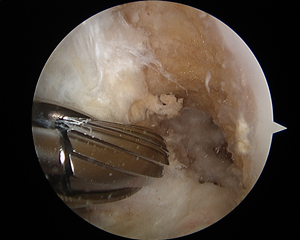
Femoral tunnel or socket for docking of the new ACL BTB graft, allowing bone-to-bone healing
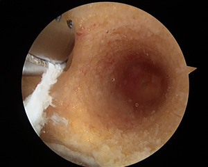
Metal tap to prepare a tract for the biocomposite interference screw
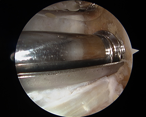
Placement of biocomposite interference screw (analogous to synthetic bone, absorbs into the bone over time with no permanent implant)
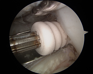
BTB graft fixed with biocomposite screw on femoral socket
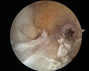
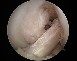
Completed ACL BTB autograft reconstruction with anatomic position and excellent quality graft tissue
Case Example #2
Notchplasty (before and after) to widen intercondylar notch tunnel and prevent graft impingement
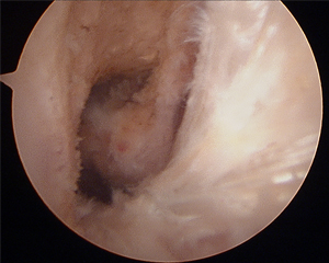
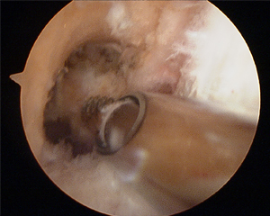
Femoral tunnel or socket for docking of the new ACL BTB graft, allowing bone-to-bone healing
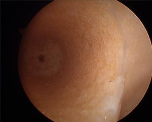
Guide pin to set femoral tunnel location
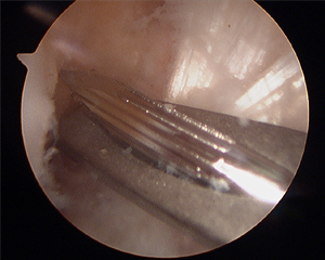
Tibial tunnel for ACL BTB graft
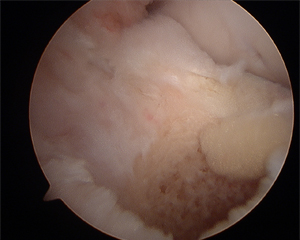
Placement of biocomposite interference screw (analogous to synthetic bone, absorbs into the bone over time with no permanent implant)
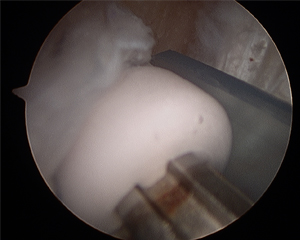
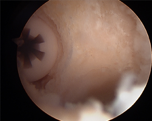
Completed ACL BTB autograft reconstruction with anatomic position and excellent quality graft tissue
nodisplay
nodisplay
Knee Procedures
- ACL Reconstruction
- PCL Reconstruction
- MCL Reconstruction
- LCL Repair And Reconstruction
- Anterolateral Ligament Reconstruction
- All-inside Meniscus Repair
- Menisectomy
- Tibial Tubercle Osteotomy
- Arthroscopic Lateral Release
- Plica Excision
- Loose Body Removal
- Synovectomy
- Infrapatellar Fat Pad Debridement
- Subchondroplasty
- Chondroplasty
- Quadriceps Tendon Repair
- Patellar Tendon Repair
- Total Knee Arthroplasty
- Medial Patellofemoral ligament (MPFL) reconstruction
- Cartilage Procedures: Microfracture, Drilling, Osteochondral Autograft Transfer, Osteochondral Allograft Transplant
- Platelet Rich Plasma


