Dr. Rice has joined Beacon Orthopedics and Sports Medicine
Congratulations Dr. Rice: 2025 Cincinnati Magazine Top Doctor
Services
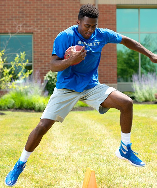
BOOST Functional Sport Training Program
BOOST Functional Sport Training is a program aimed to progress an athlete’s ability to safely return to sport. This may include return to sport from an injury treated either nonsurgically (i.e. Knee MCL sprain) or surgically (i.e. Knee ACL tear and reconstruction, Shoulder dislocation and labral repair).
Sporting activities like running, jumping and cutting demand a high level of strength and neuromuscular control, not only from the legs but from the hips, gluts and core, and similar principles apply to the upper body.
Mastering these skills is typically not within the scope and timeframe of physical therapy services, but is vital to a safe and successful integration back into sports. Whereas many patients are satisfied returning to normal activities of daily living, BOOST Functional Sport Training was developed and designed with high-level, competitive athletes in mind. This is a population with greater physiologic demands and require a more rigorous program to prepare them for the competitive field of play.
BOOST picks up where physical therapy leaves off and addresses the areas where athletes likely have post-operative deficits to minimize the risk of re-injury during play. This program typically unfolds in months 4-6 after recovery of more intensive injuries and surgeries, such as ACL reconstruction, shoulder labral repair, or hip arthroscopic labral repair. Team trainers and other specialists then typically continue care around 6 months, after functional sport training is complete and the athlete has passed all return-to-sport testing.”
BOOST Injury Prevention Programs
BOOST injury prevention program is designed to reduce injury incidence in competitive athletes. Extensive research with protocols from NFL, NCAA, FIFA, Cincinnati Reds, and other organizations have produced this comprehensive BOOST program. Research to date indicates this program reduces the incidence of ACL tears, hamstring tears, ankles sprains, and other common injuries by 30-50%! The goal is to keep athletes on the field so they can reach their full potential. BOOST injury prevention comprises a 10-20 minute dynamic warmup/workout to precede practices and games, with separate preseason, in-season, and gameday programs. Initially the athletic trainers will help teach and implement the programs, with continued supervision and support of the athletes by the coaches. Most of the stretches and exercises will be familiar to athletes, and the trainers will teach any that are unclear or unfamiliar. This program is free. There are no costs for the athletes at our participating TriHealth-affiliated high schools and colleges. Please see attached a long format guide describing the stretches and exercises in detail, as well as program rationale.
X-rays
X-rays are waves of electromagnetic radiation that can penetrate through objects of low density. They are used in a branch of medicine called radiology to create images of the inner structures of the body such as the bones and organs. X-rays are one of the most common radiology procedures and are usually the first imaging tests to be performed.
How it works
The human body is composed of tissues and organs of varying densities. X-rays directed at the body may get absorbed, reflected or pass through the different structures depending on their densities. X-rays that pass-through structures such as bones appear white on film or other media. Less dense tissues, through which X-rays may pass, create shades of grey on the image.
Indications
X-rays may be performed to look for abnormalities in bone and soft tissues that may be causing certain symptoms. They may help identify and evaluate fractures, pneumonia, cancer, intestinal obstruction, air or fluid collection, and the position of instruments or implants during a procedure or surgery.
Procedure
X-rays are usually performed by a radiologist or X-ray technician. You may be instructed to remove jewelry or metal objects that may interfere with the test or results.
The examiner positions you according to the area that needs imaging. The X-ray beam is then directed across the area. You will have to remain still during the procedure and may be instructed to hold your breath. X-rays may be repeated or taken from different angles for clarity.
A contrast dye which is visible on X-rays may sometimes be injected or swallowed before or during the procedure to help improve the clarity of certain areas.
Once the test is complete, you can resume your regular activities. Your radiologist and doctor will review the results and discuss the findings with you.
Risks and complications
X-ray imaging is a safe diagnostic tool that is extensively used to detect many conditions and abnormalities. Small amounts of radiation are used during X-rays. It may carry a small risk of affecting a developing fetus and therefore must be avoided during pregnancy.
Side effects are uncommon when a dye is used to improve the image quality, but may include allergic reaction, nausea, hives, itching and lightheadedness.
MRI
MRI or Magnetic Resonance Imaging scan is an imaging test that creates pictures of internal body structures (bones and soft tissues) with the help of magnetic fields and radio waves
The MRI can also be combined with other imaging techniques to provide a more definitive diagnosis. The scan is often used to clarify findings from previous X-rays or CT scans.
Indications
MRI scans provide information on a variety of conditions and procedures and to assess function of the internal organs such as:
- Brain and spinal cord abnormalities
- Prostate, liver and breast abnormalities
- Function and structure of the heart
- Joint problems
- Blood flow through blood vessels
- chemical composition of tissues
- Tumor detection and help in staging (tumor size, severity and spread)
Magnetic Resonance Imaging (MRI)
Procedure
Before the procedure, you will be asked to remove any metallic devices such as hearing aids, hairpins, removable dental work or other objects that may interfere with the procedure. You may be provided with ear plugs or music to block the strong noises from the MRI scan. You may be sedated if required.
The MRI machine consists of a large strong magnet and a table that moves into the opening of the scanner. During the procedure, you will be asked to lie on the table, which will be advanced into the scanner. The machine creates a magnetic field that creates loud noises. In some cases, a contrast dye may be injected through your arm to provide a clearer view of the scan. A radio wave antenna directs signals to the body and receives them back to create images by a computer attached to the scanner. You need to keep very still throughout the scan as movement may blur the resulting images. The entire procedure may take up to an hour to complete.
If you were not sedated, you may resume your usual activities immediately after the MRI. If you have been given a sedative, you will need to arrange for a relative or friend to take you home after the scan.
Advantages & Disadvantages
Advantages of MRI include:
- Does not use radiation
- Is noninvasive
- Can take images of any part of the body from almost any direction and orientation
- Produces better images of soft-tissue structures compared to other imaging techniques
- Can differentiate between tissues based on their biochemical properties such as water, fat, iron
- Can scan large regions of the body
Disadvantages of MRI include:
- Certain patients who get nervous in small spaces (claustrophobic) may not be able to have an MRI.
- Elderly or ill patients may find it difficult to cooperate, which may result in blurred images.
- MRI cannot be done on patients with implanted medical devices such as aneurysm clips in the brain, heart pacemakers and cochlear (inner ear) implants
- MRI is an expensive procedure
Risks and complications
Since an MRI scan is a noninvasive test, it is a very safe procedure. However, there is a very small risk of an allergic reaction to the contrast dye or sedation medicine if used. Any metal or electronic devices in your body are a safety threat and you should not undergo an MRI in those circumstances. Before your MRI test, make sure you notify your doctor and the MRI technologist if you:
- Have any health conditions, such as kidney or liver problems that may prevent you from having an MRI using contrast material
- Are pregnant as the effects of magnetic fields on the baby are not yet known
Bracing
Sports activities play a crucial role in maintaining a healthy body structure of an individual. However, sports activities can result in various injuries that require adequate care. If neglected, sports injuries may induce serious complications and may even require surgery of the affected area. Braces are widely recommended for the management of such sports injuries. Bracing plays an important role, both in prevention and therapeutic management of the sports injuries.
Braces or splints are specifically designed devices that safely support the injured area in correct position to induce the healing process. Braces are comprised of specific material that provides warmth and compression to the affected joint and soft tissues. Bracing involves wrapping the specific brace or splint around the injured area as recommended by your physician. Bracing helps to prevent further damage of the injured area, without affecting the daily activity of the person. Braces also are effective in healing muscular injuries, including damage to soft tissues such as tendons and ligaments.
The type of bracing depends upon the affected area (knee, wrist, and elbow etc.) and severity of the sports injury. Usually bracing will be more effective in treating mild to moderate injuries, rather than abrupt severe injuries.
For better recovery of the sports injury, bracing should be used along with physical therapy and strengthening exercises.
Orthopedic Conditions and Procedures


